Microscopy Guide
1. Choosing Your Microscope
Selecting the right microscope depends on what you want to observe. Here's a breakdown to help you pick the perfect tool for your application, along with some example models from ZEISS.
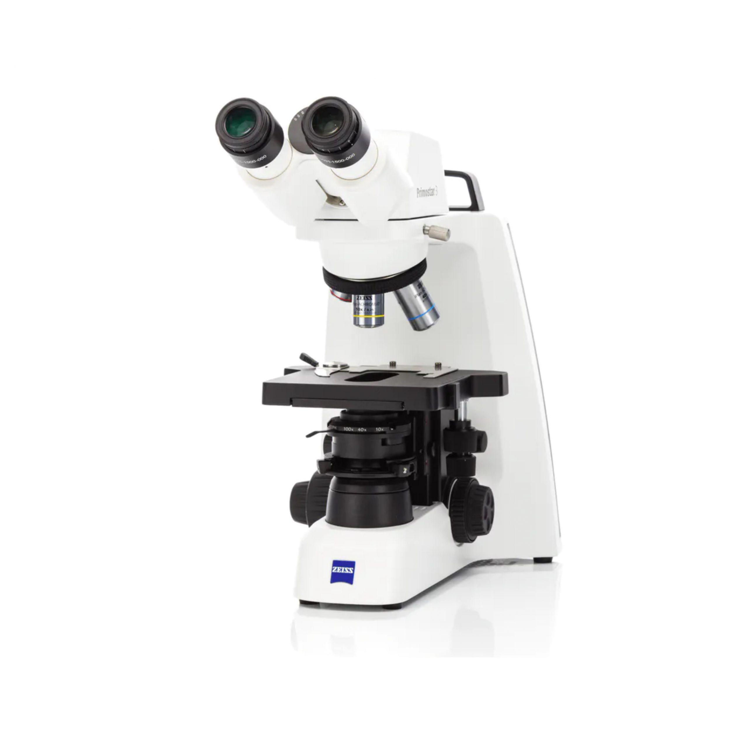 🔬 Biological Microscope: Ideal for live cell imaging, tissue sections, bacteria, and blood samples.
 These microscopes use transmitted light and high magnification to reveal structures inside thin, transparent samples.
Often paired with stains or contrast techniques. Example ZEISS models: Axio Lab.A1, Primostar 3
 ✨ Fluorescence Microscope: Ideal for targeted molecular labeling, protein tracking, and immunofluorescence.
Uses high-intensity light to excite fluorescent dyes or proteins in a sample. Great for visualizing specific structures like DNA or organelles.
Example ZEISS models: Axio Imager 2, Axio Observer
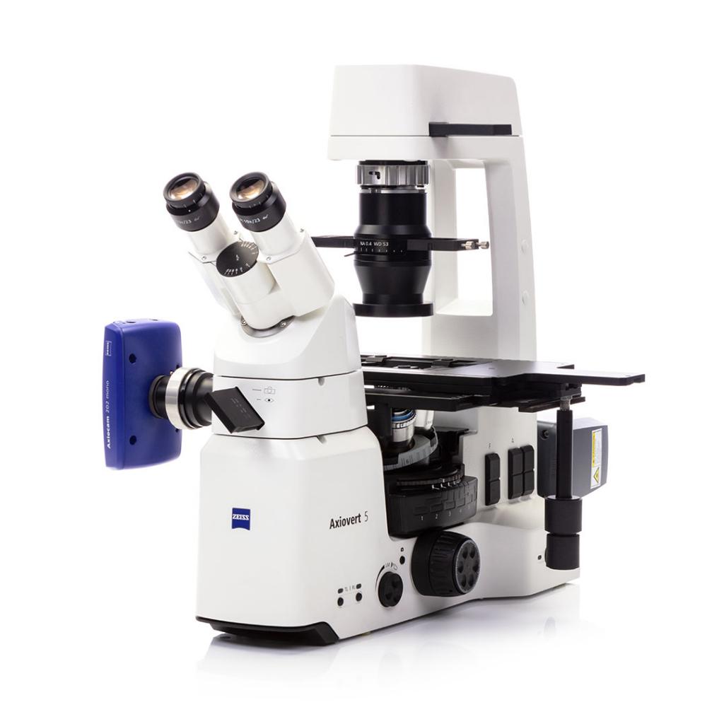
 🛠️ Metallurgical Microscope: Ideal for surface inspection, metallurgy, and semiconductor analysis.
Designed for reflected light microscopy on opaque materials like metals and composites.
Example ZEISS models: Axio Scope.A1 MET, Axiovert 5 MAT
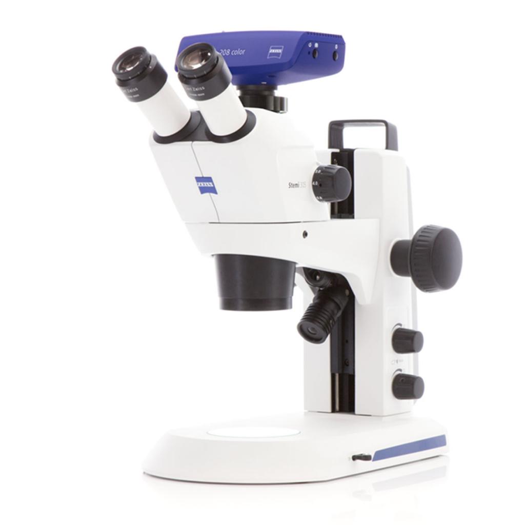 🧠 Stereomicroscope (Dissecting Microscope): Ideal for 3D observation, electronics inspection, and dissection.
Offers a wider field of view and depth perception at lower magnification.
Example ZEISS models: Stemi 305, Stemi 508
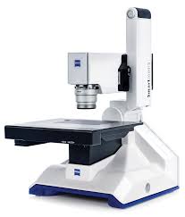 📷 Digital Microscope: Ideal for documentation, remote collaboration, and training environments.
Equipped with cameras and software for measurement and image sharing. Example ZEISS models: Smartzoom 5, Axiocam-equipped systems
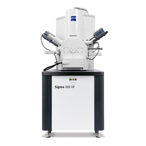 ⚛️ Electron Microscope (SEM/TEM): Ideal for nanostructures, viruses, and ultra-high-resolution imaging.
Uses electron beams for incredibly detailed views of sample surfaces or internal structures.
Example ZEISS models: Sigma 300 (SEM), Libra 200 (TEM)
2. Application Notes & Technical Tips
Fluorescence Imaging with Axiocam 807 Mono
- Use low excitation to reduce phototoxicity
- Use cooling to minimize noise
- Use flat-field correction for illumination uniformity
Image Stitching
- Use motorized stage and 10-15% field overlap
- Use software stitching with shading correction
Live Cell Imaging
- Maintain 37°C, 5% CO₂
- Minimize exposure during time-lapse
Maintenance & Calibration
- Clean optics with proper kits
- Calibrate using micrometer slide
Troubleshooting
- Noise: Lower gain or cool sensor
- Lag: Close apps or reduce resolution
3. Advanced Microscopy Techniques
Phase Contrast
Enhances contrast in transparent specimens without staining. Best for live cells.
DIC (Nomarski)
Provides pseudo-3D relief images. Useful for detailed organelle visualization.
Darkfield
Highlights edges and particles in thin, transparent specimens via scattered light.
Multiphoton
Infrared-based deep tissue fluorescence with reduced phototoxicity.
SPM (AFM / STM)
Surface scanning for nanotechnology and materials research.
Portable / Handheld
Forensics, field inspection, or rapid low-mag documentation.